The Ultimate Guide to Feline and Canine Dermatology: Causes, Clinical Signs, Treatment, and Prevention
2 months ago
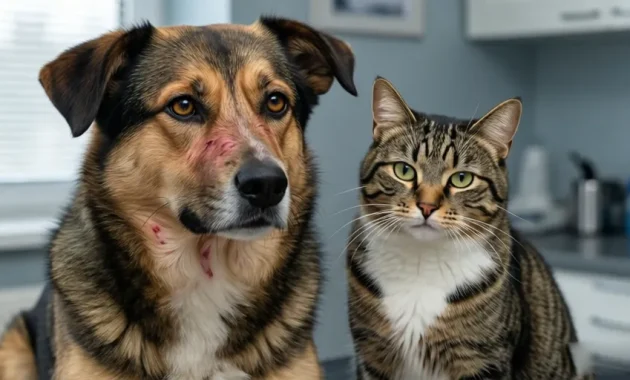
Discovering a bald patch, an angry red rash, or listening to the non-stop thump-thump-thump of your dog or cat scratching their ears in the middle of the night is a universal experience for pet parents. Skin disorders are among the most frequent reasons domestic pets visit veterinary clinics worldwide.
Because the skin is a highly visible, external organ system, dermatological issues quickly impact a pet’s quality of life. The constant itching, licking, biting, and scratching caused by skin irritation can turn a happy pet into a restless, anxious, and exhausted animal.
Understanding the root causes of these conditions—ranging from microscopic mites and fungal spores to environmental allergies and hormonal imbalances—is essential for any dedicated pet owner. This comprehensive guide breaks down the science of veterinary dermatology, explores the primary causes of skin issues, describes specific clinical symptoms, and details professional treatment protocols and preventative measures to keep your pet’s skin and coat healthy.
The Anatomy and Physiology of Pet Skin

To understand why skin diseases manifest so dramatically, we must first look at the unique biological structure of canine and feline skin. The skin is the largest organ of the body, serving as a dynamic, protective shield against environmental pathogens, mechanical trauma, and chemical irritants.
[ Epidermis: Cellular Shield ] -> [ Dermis: Hair Follicles, Glands, Blood Vessels ] -> [ Hypodermis: Subcutaneous Fat ]
-
The Epidermal Barrier: The outermost layer, the epidermis, is a shifting ecosystem of cells that continuously renews itself. In dogs and cats, the epidermis is remarkably thin compared to human skin—often only 3 to 5 cells thick (humans possess 10 to 15 layers). This thinness makes our pets highly vulnerable to superficial chemical irritants, abrasions, and localized infections.
-
The Micro-Climate of the Dermis: Beneath the epidermis lies the dermis, a complex matrix containing blood vessels, nerves, hair follicles, sebaceous (oil) glands, and apocrine (sweat) glands. Unlike humans, dogs and cats do not sweat through their skin to regulate temperature (except through their paw pads). Instead, their skin glands focus heavily on producing sebum, an oily secretion that coats the hair shaft, conditions the skin, and forms an antimicrobial lipid barrier.
-
The Follicular Ecosystem: Cats and dogs possess compound hair follicles, meaning multiple hair shafts emerge from a single pore. When inflammation strikes a single follicle, it can compromise a significant cluster of hair, leading to rapid, patchy baldness.
When any variable alters this delicate ecosystem—whether it is a surge in hormones, an overgrowth of microflora, or the physical tunneling of a parasite—the skin's defensive barrier collapses. This structural breakdown triggers an immune response that manifests as inflammation, redness, and itching.
Recognizing the Signs: The Feline and Canine Dermatological Checklist
Pets cannot tell us when their skin feels tight, burning, or intensely itchy. Instead, they communicate their physical discomfort through specific behavioral shifts and physical lesions. As a pet owner, you must learn to read these subtle and overt signs of cutaneous distress.
Behavioral Red Flags
-
Compulsive Pruritus: Chronic, repetitive scratching using the hind legs, directed at the neck, armpits, flanks, or ears.
-
Obsessive Grooming and Licking: Directing intense licking or chewing at specific body parts, particularly the paws, carpal joints, inner thighs, and perianal region. Feline grooming can be so intense that they physically strip the fur from their bodies (psychogenic alopecia or parasitic pruritus).
-
Head Shaking and Ear Scratching: Frequent, violent shaking of the head or tilting the head to one side, indicating deep discomfort within the external ear canal.
-
Rubbing Against Surfaces: Pressing and rubbing the muzzle, face, or back against carpets, furniture, or walls to alleviate hard-to-reach itching.
Physical Cutaneous Lesions
-
Eritema (Redness): Diffuse or localized blushing of the skin, indicating increased blood flow due to active inflammation or infection.
-
Desquamation (Dandruff/Flakes): Excessive shedding of dead epidermal skin cells, appearing as dry white flakes or oily, yellow scales along the spine and rump.
-
Alopesia (Hair Loss): Can present as symmetric baldness, patchy "moth-eaten" spots, or complete denuding of large structural areas.
-
Pustules and Papules: Small, raised red bumps (papules) or fluid-filled, pimple-like lesions containing yellow pus (pustules).
-
Excoriations and Ulcers: Raw, weeping, bleeding wounds caused by self-trauma when the pet scratches or bites through their own skin.
-
Lichenification (Thickening): Chronically irritated skin that becomes thick, leathery, wrinkled, and dark gray or black due to chronic friction and inflammation.
-
Malodor (Odor): A pungent, sweet, or yeasty smell emanating from the skin or ears, confirming an overgrowth of opportunistic microorganisms.
Etiology: The Major Causes of Skin Disease
Veterinary dermatologists classify skin diseases into five primary categories based on the underlying causative agent or trigger mechanism.
                  [ Dermatological Triggers ]
                              |
     +---------+----------+---+---+----------+---------+
     |         |          |       |          |         |
[ Parasitic ] [ Bacterial ] [ Fungal ] [ Allergic ] [ Metabolic / Hormonal ]
Parasitic Dermatitis (Ektoparasit)
External parasites are the most common cause of acute and chronic skin disease in companion animals. These bugs live on, bite into, or burrow inside the skin tissue.
A. Scabies (Sarcoptic & Notoedric Mange)
Scabies is a highly contagious, intensely painful skin disease caused by microscopic, eight-legged arachnid mites. In dogs, the culprit is Sarcoptes scabiei; in cats, it is Notoedres cati.
-
Pathology: Fertilized female mites use specialized, scissor-like mouthparts to cut into the epidermis. They chew long, winding tunnels deep inside the living tissue, moving forward a few millimeters every day, feeding on cellular fluids, and leaving a trail of eggs and fecal pellets behind them.
-
Clinical Presentation: The physical presence of the mites and their metabolic waste triggers a massive, explosive hypersensitivity reaction. This causes severe, unyielding pruritus.
-
Dogs: Lesions typically emerge first on the ear margins, elbows, hocks, and ventral abdomen. The skin becomes intensely red, raw, and loses all hair.
-
Cats: Notoedres focuses heavily on the face, eyelids, and neck, creating thick, gray-yellow, concrete-like crusts that can mechanically seal the eyes shut.
-
Mite burrows into skin -> Excretes feces/eggs -> Explosive allergic reaction -> Deep crusting & hair loss
B. Demodex (Demodicitosis)
Unlike scabies mites, which are caught from other animals, Demodex mites (Demodex canis in dogs, Demodex cati in cats) are normal residents of the skin. They live in tiny, harmless numbers inside the hair follicles and sebaceous glands, passed from mother to offspring during nursing.
-
Pathology: Demodex only becomes a disease when a pet’s immune system fails to keep the mite population in check. This can occur in young puppies with developing immune systems, or in adult pets suffering from underlying conditions like Feline Leukemia Virus (FeLV), Feline Immunodeficiency Virus (FIV), diabetes, Cushing's disease, or cancer.
-
Clinical Presentation: As the mites multiply into the thousands, they physically stretch and rupture the hair follicle. This leads to patchy hair loss, red or bluish-gray skin (hence the name "red mange"), a distinctive rancid odor, and the formation of deep pustules filled with blood and nanah (pus).
C. Fleas, Ticks, and Ear Mites
-
Fleas (Ctenocephalides felis): Fleas move quickly through the fur, biting the skin to consume blood meals. Their movement and biting cause localized irritation, scabbing, and general restlessness.
-
Ticks (Caplak): Ticks anchor themselves into the skin with specialized mouthparts, engorging on blood. They cause localized swelling, hard nodules, and can transmit dangerous systemic blood-borne pathogens.
-
Ear Mites (Otodectes cynotis): These surface mites inhabit the external ear canal. They bite and feed on epithelial debris, triggering intense inflammation, head shaking, and the production of a dark, dry, crumbly ear discharge that looks exactly like coffee grounds.
Infectious Dermatitis: Bacterial and Fungal Invaders
When the skin's protective lipid barrier is damaged by scratching or chronic moisture, environmental bacteria and fungi can invade the tissue, turning a mild irritation into a deep, systemic infection.
A. Impetigo (Superficial Bacterial Infection)
Impetigo is a distinct superficial bacterial infection (typically caused by Staphylococcus pseudintermedius) that primarily targets young puppies and kittens.
-
Pathology: It is closely linked to poor environmental hygiene, internal parasites, malnutrition, or a developing immune system that cannot fend off normal environmental bacteria.
-
Clinical Presentation: It manifests as a sudden outbreak of small, red bumps and delicate, pus-filled blisters (pustules) localized on the hairless skin of the abdomen, groin, and around the genitalia. These blisters are fragile; they rupture easily, leaving behind circular, peeling edges of skin (epidermal collarettes) and thin, honey-colored crusts that resemble human acne. Fortunately, impetigo is rarely deeply itchy or painful, but it indicates a breakdown in the animal's overall health defenses.
B. Ringworm (Dermatofitosis)
Despite its confusing name, ringworm is not caused by a worm. It is a highly contagious, superficial fungal infection of the hair shafts and superficial skin layers, primarily caused by the fungal species Microsporum canis, Trichophyton, or Epidermophyton.
Fungal Spore Lands on Hair -> Secretes Keratinase Enzyme -> Digests Hair Shaft -> Hair Snaps at Base -> Circular Bald Patch
-
Pathology: Fungal spores land on the skin and target keratin, the tough structural protein that makes up hair and the outer skin layers. The fungus secretes a specialized enzyme called keratinase, which digests the hair shaft from the inside out. This structurally weakens the hair, causing it to snap off right at the skin line.
-
Clinical Presentation: Ringworm produces classic, expanding circular or ring-like patches of hair loss (alopesia) with raised, red, scaly edges and a clearing center. The skin within the ring often appears dry, flaky, and ash-gray.
-
The Zoonotic Threat: Ringworm is a highly infectious zoonosis. The microscopic fungal spores can easily pass from infected dogs and cats directly to humans, especially children, the elderly, or anyone with a compromised immune system. Touching an infected pet can cause ringworm lesions on your own arms, chest, or face. It also contaminates your living space, as shed spores can survive on carpets, furniture, and grooming brushes for up to 18 months.
Allergic Dermatitis: The Hyper-Reactive Immune System
Allergic skin disease occurs when a pet's immune system overreacts to completely harmless environmental or dietary substances.
A. Flea Allergy Dermatitis (FAD)
FAD is the most common allergy diagnosed in companion animals. Pets with FAD are not just annoyed by fleas; they are profoundly allergic to specific amino acids and anticoagulant enzymes present in flea saliva.
When a flea bites, it injects a tiny drop of saliva into the dermis. In an allergic pet, a single flea bite can trigger an intense, full-body allergic reaction. The pet will lick, bite, and chew their skin until it is raw. In dogs, this typically creates a "v-shaped" pattern of hair loss and red crusts along the lower back, tail base, and thighs. In cats, it causes tiny, sand-like scabs across the neck and back, a condition known as miliary dermatitis.
B. Environmental Allergies (Atopic Dermatitis / Atopy)
Atopic dermatitis is a chronic, genetically inherited allergic skin disease where pets become hypersensitive to common environmental allergens:
-
Tree, grass, and weed pollens.
-
House dust mites and storage mites.
-
Mold spores and dander.
Pets can absorb these allergens directly across a defective skin barrier or inhale them. Atopy targets specific zones on the body. Atopic dogs and cats typically present with intense itching focused on the face, muzzle, eyes, armpits (axillae), groin, and paws. They will lick their paws continuously, causing the fur to turn a rusty brown color due to staining from porphyrin pigments in their saliva.
C. Food Allergies
Food allergies represent a reliable immunological reaction to a specific ingredient within their regular diet—almost always a primary protein source like beef, chicken, dairy, lamb, or fish.
Unlike environmental allergies, which may flare up seasonally in the spring or fall, food allergies cause year-round, non-seasonal pruritus. In addition to severe skin itching around the face, ears, and belly, food-allergic pets often suffer from concurrent gastrointestinal signs, such as chronic soft stools, flatulence, or intermittent vomiting.
Species-Specific and Complex Conditions
Some dermatological conditions are unique to a specific species due to their unique physiological layout or glandular structure.
Stud Tail (Tail Gland Hyperplasia)
Stud tail is a unique dermatological disorder seen in felines. It is caused by the structural hypertrophy and hyperactivity of the supracaudal gland cluster located along the top surface of the tail base.
Hormonal Surge -> Hyperactive Supracaudal Glands -> Excessive Sebum Production -> Matted, Waxy Fur -> Bacterial Infection
-
Pathology: The supracaudal gland is a large collection of sebaceous glands that produce waxy sebum used for territorial scent-marking. When a male cat hits puberty, circulating testosterone levels can cause these glands to expand and produce an excessive volume of oily sebum. While it is most common in intact (unneutered) adult male cats, it can also happen in neutered males and females due to individual genetic variations.
-
Clinical Presentation: The excessive, waxy oil accumulation causes the fur at the base of the tail to look permanently greasy, yellow, clumped, and matted. As the condition progresses, the hair follicles become plugged with waxy blackheads (comedones), the hair falls out, and the area develops a rancid, oily smell. If environmental bacteria invade the plugged follicles, the tail base can become swollen, red, and painful, oozing pus when pressed.
Cutaneous Masses: Abscesses, Tumors, and Cancers
Any new lump, bump, or swelling on your pet's skin warrants close professional attention. These masses are generally categorized into three types:
-
Abscesses: A localized pocket of pus beneath the skin, usually caused by a penetrating bite wound or scratch from another animal. They are hot, painful, swell rapidly, and can cause a sudden fever.
-
Benign Tumors: Non-cancerous growths such as lipomas (fatty lumps), papillomas (warts), or sebaceous cysts. These grow slowly, do not spread to other organs, and rarely cause pain unless they grow large enough to restrict movement.
-
Malignant Cancers: Dangerous growths such as Mast Cell Tumors (MCT), Squamous Cell Carcinoma, or Fibrosarcoma. These masses can look completely innocent—resembling a tiny pimple or a harmless bug bite—but they can spread silently to local lymph nodes and internal organs.
Professional Veterinary Treatment Matrix
Treating skin disease requires a precise, diagnostic approach. Attempting to treat a skin issue at home without a diagnosis can hide symptoms, make the disease worse, or accidentally poison your pet.
Diagnostic Testing: The Foundation of Therapy
-
Skin Scrapings: The vet uses a dull scalpel blade to gently scrape the skin until a tiny amount of capillary blood appears. This collects mites like Demodex or Sarcoptes from deep inside the follicles and tissue layers so they can be viewed under a microscope.
-
Fecal Flotation: Used alongside skin scrapings because pets often swallow skin mites or parasite eggs while chewing at their itchy skin.
-
Fungal Culture (DTM): Hair shafts are placed into a specialized growth medium to confirm a ringworm diagnosis. If dermatophytes are present, the gel changes color from yellow to bright red.
-
Fine Needle Aspiration (FNA) and Biopsy: A small needle is inserted into a lump to collect cells, or a small tissue sample is surgically removed, to determine if a mass is a benign cyst, an abscess, or a malignant cancer.
Targeted Medical Therapies
[ Allergic Inflammatory Cycle ] -> [ Corticosteroids / Targeted Biologics ] -> [ Block Pruritus Pathway ] -> [ Skin Healing ]
-
Corticosteroids (Prednisolone/Dexamethasone): Powerful anti-inflammatory drugs used to break the intense cycle of itching and scratching in severe allergic flare-ups. While highly effective at providing immediate relief, long-term use can cause side effects like increased thirst, frequent urination, muscle wasting, and a suppressed immune system. They should always be used at the lowest effective dose for the shortest time possible.
-
Targeted Modern Biologics and Immunomodulators: Modern veterinary medicine utilizes highly targeted, non-steroidal therapies to manage chronic allergies without the side effects of steroids:
-
Oclacitinib (Apoquel): A daily oral tablet for dogs that selectively blocks the Janus Kinase (JAK) enzyme pathway, safely stopping the itch signal before it reaches the brain.
-
Lokivetmab (Cytopoint): An injectable monoclonal antibody for dogs that mimics natural immune proteins to target and neutralize the specific cytokine (IL-31) that triggers allergic itching. A single injection can provide relief for 4 to 8 weeks.
-
Cyclosporine (Atopica): An immunomodulator safe for both dogs and cats that calms an overactive immune system, making it ideal for chronic atopic dermatitis.
-
-
Antibiotics and Antifungals:
-
Superficial bacterial infections (like impetigo) are treated with topical antibiotic ointments or oral antibiotics.
-
Fungal infections (like ringworm) require topical antifungal creams, specialized medicated baths, or oral systemic antifungal medications (like Itraconazole) for severe or widespread cases.
-
-
Specific Therapy for Stud Tail: Managing stud tail requires a multi-step approach:
-
Hormonal Control via Castration: For intact male cats, surgical castration (neutering) is the most effective choice. Removing the testicles eliminates the main source of testosterone, shutting down the hormonal drive causing the glands to overproduce oil.
-
Topical Degreasing: Washing the tail regularly with specialized antiseboroic shampoos containing benzoyl peroxide or salicylic acid to break down and flush away the heavy, waxy grease buildup.
-
Comprehensive Preventive Protocols
Preventing skin disease is significantly easier, less expensive, and more compassionate than treating a severe, long-standing dermatological breakdown.
1. Environmental Hygiene and Home Sanitation
-
Regular Laundering: Wash your pet’s bedding, crate mats, and favorite blankets weekly in hot water ($>60^\circ\text{C}$) to destroy parasite eggs, environmental mite populations, and fungal spores.
-
Strategic Vacuuming: Vacuum rugs, carpets, and upholstered furniture frequently, focusing on dark corners and baseboards where flea larvae and dust mites thrive. Always empty the vacuum bag or canister immediately into an outdoor trash bin.
2. Routine Grooming and Physical Inspections
-
Medicated and Maintenance Bathing: Bathe your dog regularly with a gentle, hypoallergenic, soap-free shampoo to wash away trapped environmental pollens and allergens before they can cross the skin barrier. Cats generally do not need regular baths due to their self-grooming habits, but regular brushing is essential to prevent painful mats and check for underlying skin issues.
-
The Flea Comb Check: Run a fine-toothed metal flea comb through your pet’s fur down to the skin level, checking for adult fleas or tiny black specks of flea dirt.
3. High-Quality Nutrition and Fatty Acid Supplementation
A healthy coat starts from within. Feed your pet a high-quality, biologically appropriate diet rich in clean animal proteins. Look for diets fortified with Omega-3 and Omega-6 essential fatty acids (sourced from high-quality fish oil or flaxseed). These fatty acids act as natural anti-inflammatories within the dermis, reinforcing the structural lipid barrier of the skin and reducing overall skin reactivity to environmental allergens.
Summary Reference Matrix
| Condition | Primary Cause | Classic Symptoms | Primary Treatment Options | Prevention Strategy |
| Scabies | Microscopic Mites (Sarcoptes/Notoedres) | Extreme itching, leathery skin, thick gray-yellow crusts on face or ears. | Antiparasitic injections (Ivermectin) or prescription topical spot-ons. | Prevent contact with stray animals; use routine monthly parasite controls. |
| Demodex | Follicular Mite Overgrowth (Demodex) | Patchy hair loss, skin redness/darkening, bleeding pustules, sour odor. | Specialized medicated dips, topical therapies, or oral antiparasitics. | Maintain strong immune health; screen for underlying systemic diseases. |
| Impetigo | Superficial Bacterial Infection (Staphylococcus) | Red bumps, pus-filled blisters on the hairless belly/groin of young puppies. | Topical antibiotic ointments or short-term oral antibiotics. | Improve environmental sanitation; provide optimal nutrition. |
| Ringworm | Superficial Fungus (Microsporum canis) | Expanding circular bald patches, dry ash-gray flakes, raised red edges. | Topical antifungal creams, medicated shampoos, or oral antifungals. | Zoonotic warning: Wear gloves; disinfect home; isolate infected pets. |
| Atopic Dermatitis | Environmental Allergies (Pollen, Dust Mites) | Intense licking of paws, face rubbing, red groin/armpits, chronic ear infections. | Target-specific biologics (Apoquel/Cytopoint), steroids, or allergen avoidance. | Regular bathing to wash away surface pollens; use fatty acid supplements. |
| Stud Tail | Hyperactive Tail Glands (Testosterone-driven) | Greasy, matted, yellow fur at the top base of the tail; blackheads; oily odor. | Surgical castration (neutering) for intact males; benzoyl peroxide shampoos. | Early neutering of male cats; regular grooming of the tail base. |
| Skin Masses | Abscesses, Cysts, Tumors, or Cancer | Visible lumps under or on the skin, localized swelling, changing skin colors. | Surgical biopsy or FNA diagnosis; surgical removal; antibiotics for abscesses. | Perform weekly full-body touch checks; seek immediate veterinary evaluation. |
Managing your pet’s skin health requires consistency, observation, and patience. Skin cells take several weeks to regenerate, meaning healing is a gradual process. By monitoring your pet's behavior, maintaining regular grooming routines, using year-round parasite prevention, and working closely with your veterinarian at the first sign of an issue, you can protect your pet from the discomfort of skin disease and keep their coat healthy and beautiful.
FAQ
Q1: What are the most common skin diseases in dogs and cats?
A: The most common skin diseases in pets include flea infestations, flea allergy dermatitis (FAD), scabies (mange), Demodex mite overgrowth, ringworm, bacterial infections such as impetigo, environmental allergies (atopic dermatitis), food allergies, ear mite infestations, and various skin masses or tumors. These conditions often cause itching, redness, hair loss, scaling, skin odor, and discomfort.
Q2: Why is my dog or cat constantly scratching and biting their skin?
A: Persistent scratching, licking, and biting are usually signs of skin irritation. Common causes include fleas, mites, allergic reactions, fungal infections, bacterial infections, dry skin, environmental allergens, and food sensitivities. Chronic scratching can lead to open wounds, secondary infections, and significant discomfort if left untreated.
Q3: How can I tell if my pet has a skin problem?
A: Early warning signs include excessive scratching, constant licking, hair loss, red skin, dandruff, scabs, foul odor, ear shaking, skin thickening, hot spots, and behavioral changes such as irritability or restlessness. Any sudden changes in your pet's coat or skin condition should be investigated promptly.
Q4: What causes hair loss in dogs and cats?
A: Hair loss can result from parasites such as fleas and mites, fungal infections like ringworm, bacterial skin infections, hormonal imbalances, allergies, excessive grooming, nutritional deficiencies, or underlying medical conditions. The pattern and location of hair loss often help veterinarians identify the underlying cause.
Q5: What is scabies in pets?
A: Scabies, also known as mange, is a highly contagious skin disease caused by microscopic mites. These mites burrow into the skin, causing severe itching, inflammation, crusting, and hair loss. In dogs, Sarcoptes mites are responsible, while cats are commonly affected by Notoedres mites.
Q6: What is the difference between scabies and Demodex mange?
A: Scabies is highly contagious and caused by mites that burrow into the skin. Demodex mites naturally live within hair follicles and only become problematic when the immune system is weakened. Scabies typically causes intense itching, while Demodex often causes patchy hair loss and skin inflammation that may or may not be itchy.
Q7: Can fleas cause serious skin disease?
A: Yes. Fleas can trigger flea allergy dermatitis (FAD), one of the most common allergic skin conditions in pets. Even a single flea bite can cause severe itching, hair loss, scabs, and skin inflammation in sensitive animals.
Q8: What are ear mites and how do I recognize them?
A: Ear mites are tiny parasites that live in the ear canal. Common signs include frequent head shaking, scratching around the ears, irritation, and a dark brown or black discharge that resembles coffee grounds. Veterinary treatment is necessary to eliminate the infestation completely.
Q9: What is ringworm and can humans catch it?
A: Ringworm is a fungal infection that affects the skin and hair. It commonly causes circular bald patches with red, scaly edges. Ringworm is zoonotic, meaning it can spread from pets to humans. Prompt diagnosis, treatment, and environmental cleaning are essential.
Q10: How do pets get ringworm?
A: Ringworm spreads through direct contact with infected animals, contaminated bedding, grooming tools, furniture, or fungal spores present in the environment. The spores can survive for many months, making environmental sanitation critical.
Q11: What is impetigo in dogs and cats?
A: Impetigo is a superficial bacterial skin infection most commonly seen in puppies and kittens. It appears as small red bumps, pustules, peeling skin, and crusts, especially on the abdomen and groin. It often develops when immunity is immature or compromised.
Q12: What is atopic dermatitis?
A: Atopic dermatitis is a chronic allergic skin disease caused by hypersensitivity to environmental allergens such as pollen, dust mites, mold spores, and dander. It often results in recurring itching, paw licking, ear infections, and skin inflammation.
Q13: How can I tell if my pet has environmental allergies?
A: Pets with environmental allergies often lick their paws excessively, rub their faces, scratch around the ears, develop chronic ear infections, and experience redness in the armpits or groin. Symptoms may worsen during specific seasons depending on the allergen.
Q14: Can food allergies cause skin problems?
A: Yes. Food allergies commonly cause year-round itching, ear inflammation, facial irritation, excessive licking, and digestive symptoms such as vomiting, diarrhea, or chronic soft stools. Common food triggers include beef, chicken, dairy products, fish, and lamb.
Q15: What is Stud Tail in cats?
A: Stud Tail is a condition involving excessive activity of the sebaceous glands located at the base of the tail. It causes greasy fur, blackheads, oily skin, hair loss, and sometimes secondary bacterial infections. It is most commonly seen in intact male cats.
Q16: Why does my pet's skin smell bad?
A: A strong odor often indicates bacterial or yeast overgrowth. Skin infections, ear infections, allergies, excessive oil production, and poor skin health can all produce unpleasant smells. Persistent odor should always be evaluated by a veterinarian.
Q17: What diagnostic tests are used for skin diseases in pets?
A: Veterinarians commonly perform skin scrapings, fungal cultures, cytology, biopsies, fine needle aspirations (FNA), allergy testing, blood work, and microscopic examinations to identify parasites, infections, allergies, or skin cancers.
Q18: How are skin allergies treated in dogs and cats?
A: Treatment depends on the cause and may include allergy management, medicated shampoos, parasite control, dietary trials, antihistamines, corticosteroids, immunomodulators, biologic therapies, and environmental modifications to reduce allergen exposure.
Q19: How can I prevent skin diseases in my pet?
A: Prevention includes year-round flea and tick control, regular grooming, routine veterinary examinations, maintaining a healthy diet rich in omega fatty acids, frequent bedding cleaning, vacuuming living spaces, and promptly addressing any signs of itching or skin irritation.
Q20: When should I take my pet to a veterinarian for a skin problem?
A: Seek veterinary attention if your pet experiences persistent itching, hair loss, open wounds, ear discharge, foul odor, skin thickening, recurring infections, unexplained lumps, or symptoms that continue for more than a few days. Early diagnosis often leads to faster and more effective treatment.


